| Cellular and Molecular Medicine Research, ISSN 2817-6359 online, Open Access |
| Article copyright, the authors; Journal compilation copyright, Cell Mol Med Res and Elmer Press Inc |
| Journal website https://cmmr.elmerpub.com |
Review
Volume 4, Number 1, September 2026, pages 1-9

Molecular Diagnostics in Barrett’s Esophagus: Bridging Endoscopy and Cellular Pathobiology to Detect Early Esophageal Adenocarcinoma
Ahmed Abdallah Salman 
Internal Medicine Department, Faculty of Medicine, Cairo University, Cairo 11562, Egypt
Manuscript submitted July 6, 2026, accepted August 17, 2026, published online September 2, 2026
Short title: Molecular Diagnostics in Barrett’s Esophagus
doi: https://doi.org/10.14740/cmmr116
| Abstract | ▴Top |
Esophageal adenocarcinoma (EAC) is among the most lethal gastrointestinal malignancies, and its incidence has risen markedly in Western populations over the last 50 years. Barrett’s esophagus (BE), the intestinal-type metaplasia that replaces the distal esophageal squamous mucosa, is the only known precursor of EAC and underpins current early-detection practice. Current surveillance—periodic endoscopy with random four-quadrant (Seattle protocol) biopsies graded for dysplasia—has two well-characterized limitations: forceps biopsies sample only a small portion of the metaplastic surface, and histopathological diagnosis of dysplasia, particularly low-grade dysplasia, has considerable interobserver variability. These limitations have prompted molecular approaches to risk stratification. This narrative review links the cellular and molecular pathobiology of the Barrett’s-to-EAC sequence with diagnostic approaches for risk stratification and early detection. We first summarize the genomic architecture of progression—TP53 loss and clonal expansion in a subset of progressing Barrett’s lesions, often preceding whole-genome doubling, together with epigenetic dysregulation and late oncogene amplification—along with evidence that BE is derived from gastric cardia progenitors at the single-cell level. We then review tissue-based adjuncts that provide objective information from the biopsy: p53 immunohistochemistry, multiplexed tissue systems pathology (the TSP-9/TissueCypher assay), and wide-area transepithelial sampling with computer-assisted three-dimensional analysis (WATS3D). We also appraise non-endoscopic molecular cell-collection devices used to triage patients for endoscopy: the Cytosponge coupled with trefoil factor 3, and the encapsulated balloon (EsoCheck) coupled with a methylated DNA panel (EsoGuard; methylated vimentin and cyclin A1). Finally, we consider liquid biopsy based on circulating cell-free DNA methylation, with artificial intelligence as a computational adjunct. Together, these modalities may convert the qualitative, observer-dependent question “Is there dysplasia?” into a quantitative assessment of an individual’s risk of progression. We examine where the evidence is strongest, where it remains preliminary, and how these tools could be combined into a risk-aligned surveillance model that detects EAC at a curable stage while reducing unnecessary procedures for patients at low risk.
Keywords: Barrett’s esophagus; Esophageal adenocarcinoma; Molecular diagnostics; DNA methylation; Risk stratification
| Introduction | ▴Top |
Esophageal adenocarcinoma (EAC) is a lethal malignancy and increasingly burdensome globally. Global estimates place esophageal cancer among the leading causes of cancer mortality, with the adenocarcinoma subtype now predominating in high-income Western countries after a multi-fold rise since the 1970s [1]. Five-year survival is poor because patients typically present with dysphagia or weight loss only after the tumor has invaded deeply or metastasized, and the single clearest opportunity to change this trajectory is to detect neoplasia at a pre-invasive or intramucosal stage, when endoscopic eradication is curative.
Barrett’s esophagus (BE)—the replacement of the normal stratified squamous lining of the distal esophagus by columnar, intestinal-type epithelium in response to chronic gastroesophageal reflux—is the only established precursor lesion for EAC and therefore the biological basis of early-detection programs [2]. Malignant transformation is represented as a stepwise metaplasia–dysplasia–adenocarcinoma sequence. The absolute risk of progression is low for any one patient: a nationwide, population-based cohort estimated the annual incidence of EAC in patients with BE at approximately 0.12%, an order of magnitude lower than the figure used historically to justify rigorous surveillance [2]. Because progression is rare, surveillance exposes a large population to repeated procedures to identify relatively few progressors. The clinical task is therefore not simply to diagnose BE, but to identify which patients are likely to progress.
Professional societies on both sides of the Atlantic recommend endoscopic surveillance of patients with BE, using high-definition white-light endoscopy with virtual chromoendoscopy and systematic four-quadrant biopsies at defined intervals—the Seattle protocol—in addition to targeted sampling of any visible lesion [3, 4]. Surveillance is based on two sequential judgments, and both have limitations. The first is spatial: random forceps biopsies sample only a small fraction of the Barrett’s surface area, so focal dysplasia or early cancer can be missed entirely. The second is interpretive: the histopathological classification of dysplasia, particularly the distinction between reactive changes and low-grade dysplasia (LGD), has considerable interobserver disagreement, and many community diagnoses of LGD are downstaged on expert review [5]. Together, these problems reduce the sensitivity and reproducibility of morphology-based surveillance and have prompted evaluation of objective, biologically based adjuncts.
These limitations have prompted development of molecular assays that characterize the cellular and genomic changes underlying metaplasia rather than morphology alone. Current guidelines reflect part of this change: the latest American College of Gastroenterology guideline expanded permissible screening to non-endoscopic methods [3], and an American Gastroenterological Association clinical practice update recognized non-endoscopic cell-collection devices, wide-area transepithelial sampling, and a tissue systems pathology prediction assay as adjuncts to conventional practice [5]. This review links the cellular origin and molecular progression of BE to the clinical evidence for each diagnostic modality. It first describes the Barrett’s-to-EAC sequence and then evaluates how tissue-based, non-endoscopic, circulating, and computational approaches may complement endoscopy-guided histology. The discussion also considers these technologies within the change from primarily surgical treatment to endoscopic and molecular management [6].
| Methods | ▴Top |
This is a narrative rather than a systematic review. PubMed/MEDLINE was searched through August 14, 2026, for English-language articles. The structured update combined “Barrett’s esophagus,” “esophageal adenocarcinoma,” “dysplasia,” “surveillance,” and “screening” with modality- and mechanism-specific terms including “TP53 mutation,” “biallelic TP53 loss,” “genome doubling,” “clonal evolution,” “epigenomics,” “DNA methylation,” “p53 immunohistochemistry,” “tissue systems pathology,” “TissueCypher,” “wide-area transepithelial sampling,” “Cytosponge,” “trefoil factor 3,” “EsoCheck,” “EsoGuard,” “cell-free DNA,” “artificial intelligence,” “diagnostic accuracy,” “cost-effectiveness,” “implementation,” “race,” “ethnicity,” and “health equity.” Reference lists of key studies, systematic reviews, and current society guidelines were also examined. Open-access full text was prioritized because of access constraints, while landmark studies, pivotal trials, systematic reviews and meta-analyses, and current guidelines were preferentially included. Each newly added reference was checked against PubMed, including its digital object identifier (DOI) and PubMed identifier (PMID). Selection was purposive rather than exhaustive, and no protocol, duplicate independent screening, formal risk-of-bias assessment, or quantitative synthesis was undertaken. Because one author performed study selection and interpretation, selection and confirmation bias remain possible.
| Cellular and Molecular Pathobiology of the Barrett’s–Adenocarcinoma Sequence | ▴Top |
The biological basis of these assays can be considered through two questions: where Barrett’s metaplasia originates and how it progresses to cancer.
Cellular origin
The cell from which BE originates has long been debated. Combining single-cell transcriptomics, methylation- and chromatin-based lineage inference, somatic mutation analysis, and organoid modeling, a global molecular-phenotyping study concluded that Barrett’s epithelium is most similar to, and most likely stems from, the gastric cardia, with the metaplastic program controlled by transcription factors such as c-MYC and HNF4A [7]. The same study also proposed that EAC might develop from undifferentiated Barrett’s cell types without any pathologically identifiable dysplastic precursor—an observation with important implications for early detection, since it indicates that some cancers will develop without necessarily passing through an easily biopsiable, morphologically obvious intermediate. This recasting of BE away from a mere histologic appearance and toward a definable cellular state is precisely the orientation of molecular assays (Fig. 1).
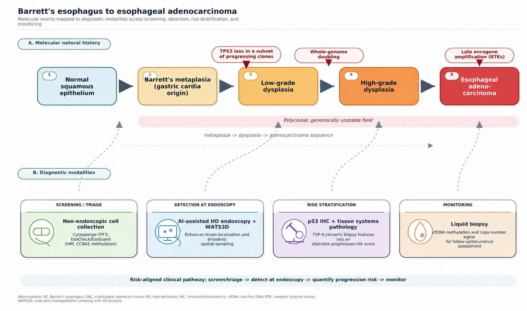 Click for large image | Figure 1. The molecular natural history of the Barrett’s esophagus-to-adenocarcinoma sequence (top: metaplasia of gastric-cardia origin, with TP53 loss and clonal expansion in a subset of progressing lineages, often before whole-genome doubling, followed by late oncogene amplification) mapped to the diagnostic modalities by stage (bottom: non-endoscopic cell collection for screening; AI-assisted endoscopy and WATS3D for detection; p53 immunohistochemistry and tissue systems pathology for risk stratification; and cell-free DNA for monitoring). |
Genomic evolution
Whole-genome sequencing of paired Barrett’s and adenocarcinoma samples revealed that BE is polyclonal and can be widely mutated even when non-dysplastic, with a mutational burden approaching that of EAC [8]. As invasion develops, copy-number alterations increase and clonal heterogeneity persists; consequently, the coding mutations in an EAC may overlap only partly with those in immediately adjacent Barrett’s mucosa [8]. This spatial diversity provides a molecular explanation for sampling error, because a forceps biopsy captures only one region of a genetically heterogeneous field. Paired-exome analyses showed that a TP53 mutation can be an early, clonally shared event in some progressing lineages, including in some non-dysplastic epithelium, whereas oncogene amplification generally occurs later [9]. This is not an obligatory sequence: TP53 alterations are absent from many stable non-dysplastic segments, and the clinically consequential event appears to be expansion of a TP53-deficient clone with structural instability rather than any TP53 mutation in isolation. In the paired-exome dataset, 62.5% of EACs emerged after whole-genome doubling, supporting a punctuated route in which TP53 loss, genome doubling, and oncogene amplification can occur in succession [9]. The omics literature adds transcriptomic, proteomic, epigenomic, and microbiome layers to this genomic framework and shows that TP53 loss, CDKN2A alteration, chromosomal instability, and epigenetic remodeling occur with variable timing and combinations [10]. EAC is characterized by copy-number alterations, a high mutational load, and frequent co-amplification of receptor tyrosine kinases, features that distinguish it from esophageal squamous carcinoma and have opened research into a putative cancer stem-cell compartment [11].
Relation to biomarkers
Molecular abnormalities can precede the morphological changes used in surveillance and may predict progression. Genomic evaluation of non-dysplastic Barrett’s tissue could identify patients at increased risk before dysplasia is recognized histologically [12]. Expansion of TP53-deficient clones, aneuploidy, epigenetic dysregulation, and clonal diversity are recurrent but nonobligate routes to progression. Assays that quantify these features may complement subjective histological grading, but this requires prospective validation. The following sections review tissue-based, non-endoscopic, and circulating approaches.
| The Limits of Endoscopy-Guided Histopathology and the First Molecular Adjunct | ▴Top |
Advanced assays should be assessed against the limitations of current surveillance because each addresses a different deficiency.
Sampling error and interobserver variability
The Seattle protocol, even when strictly adhered to, samples only a small fraction of the Barrett’s surface, and adherence in routine practice is frequently incomplete. Superimposed on this spatial problem is a diagnostic one. The grading of dysplasia is a subjective, morphology-based exercise with limited reproducibility; the diagnosis of LGD in particular is unreliable, and its predictive value for progression is weak when rendered by generalist pathologists. A rigorous histological study concluded that a panel of reproducible criteria—loss of surface maturation, mucin depletion, nuclear enlargement, and increased mitotic activity—could stratify patients with LGD into meaningfully different risk groups, and that combining these criteria with p53 immunohistochemistry improved the prediction of progression [13]. The lesson is twofold: morphology provides a genuine prognostic signal, but reliably extracting it requires either strict criteria applied by experts or an objective molecular overlay.
p53 immunohistochemistry
This widely available adjunct uses abnormal p53 expression as a surrogate for TP53 alteration. Overexpression or complete loss can be assessed on routine biopsy sections. A systematic review and meta-analysis found a strong association between aberrant p53 staining and neoplastic progression in BE, supporting its use alongside dysplasia assessment [14]. British guidance and some expert frameworks consider p53 immunohistochemistry helpful, particularly when morphology is equivocal, but major societies have not endorsed routine biomarker-directed surveillance because clinical utility remains insufficiently established [3, 4, 15]. The test is relatively inexpensive and widely available. However, it evaluates one pathway and retains the spatial sampling limitations of forceps biopsy. A negative result cannot exclude progression because TP53-driven evolution is not universal.
| Tissue Systems Pathology and Wide-Area Sampling: Objective Information From the Biopsy | ▴Top |
p53 immunohistochemistry evaluates one molecular pathway in a biopsy. Tissue systems pathology measures several features within the same specimen, whereas wide-area transepithelial sampling with computer-assisted three-dimensional analysis (WATS3D) increases the tissue area sampled.
Tissue systems pathology (the TSP-9/TissueCypher assay)
Using multiplexed immunofluorescence on a standard formalin-fixed biopsy, this method quantifies a panel of epithelial and stromal biomarkers, along with their morphometric features, and integrates them, via a validated algorithm, into an individualized progression-risk score and class (low, intermediate, high). In a retrospective analysis of the screening cohort of a randomized trial, the assay objectively risk-stratified patients with LGD and, importantly, identified a substantial fraction of true progressors among patients whom expert pathologists had downstaged to non-dysplastic BE [16]. A pooled analysis of international multicenter studies then showed that a high-risk class was an independent predictor of progression to high-grade dysplasia (HGD) or EAC, improving risk prediction over clinical variables alone, with high specificity but only moderate sensitivity [17]. Subsequent work established that the assay predicts progression independently of standard clinicopathological variables and can flag high-risk patients even within non-dysplastic BE, whose measured progression rate in the high-risk class approached that of expert-confirmed LGD [18]. In a head-to-head comparison against 30 pathologists from five countries, the test detected progressors with higher sensitivity than the average pathology review, suggesting that it can partially compensate for the variability in LGD interpretation [19]. Collectively, these studies position tissue systems pathology as a means of upstaging surveillance for genuinely high-risk patients while reassuring those at low risk—precisely the discrimination that morphology alone struggles to provide. The caveats are that sensitivity, while superior to generalist review, is incomplete, and that most of the evidence derives from retrospective cohorts.
WATS3D
Whereas tissue systems pathology extracts more information from a single biopsy, WATS3D targets the complementary problem of spatial sampling. An abrasive brush collects a broad, full-thickness (“transepithelial”) sample of the Barrett’s segment, which is then analyzed with computer-assisted three-dimensional reconstruction and neural-network-based flagging of abnormal cells for pathologist review. Used as an adjunct to Seattle-protocol forceps biopsy, WATS3D increases the detection of BE and of dysplasia: a systematic review and meta-analysis reported a meaningful absolute increase in the yield of dysplasia over forceps biopsy alone, although the authors appropriately cautioned that the natural history and clinical significance of dysplasia detected by WATS3D alone remain incompletely defined [20]. Real-world data indicate that a positive WATS3D result frequently changes management—prompting enrollment in surveillance, escalation of surveillance frequency, initiation or intensification of acid suppression, or referral for endoscopic therapy [21]. WATS3D therefore broadens the spatial reach of sampling but also introduces uncertainty about how to act on isolated WATS3D-only findings. Both tissue-based strategies share a defining feature: they still require an endoscopy. The following section turns to tools designed to remove that requirement.
| Non-Endoscopic Molecular Cell Collection: Decoupling Screening From Endoscopy | ▴Top |
The greatest structural limitation of endoscopic surveillance is that it is resource-intensive, uncomfortable, and therefore impractical as a population-screening instrument for a disease whose precursor is common but whose progression is rare. Non-endoscopic cell-collection devices, paired with molecular readouts, are engineered to solve this by capturing esophageal cells at the point of care and triaging patients to endoscopy only when a molecular signal is positive.
The Cytosponge coupled with trefoil factor 3 (TFF3)
The Cytosponge is a compressed mesh sphere within a dissolvable capsule; the patient swallows it on a string, the capsule dissolves in the stomach, and the expanded sponge is withdrawn, collecting cells along the length of the esophagus. The collected cells are stained for TFF3, a marker of intestinal metaplasia. In the pragmatic, cluster-randomized BEST3 trial conducted in primary care, offering the Cytosponge-TFF3 test to patients on long-term acid suppression increased the detection of BE roughly 10-fold compared with usual care over 12 months, and it also identified cases of dysplastic BE and early cancer that would otherwise have gone undiagnosed [22]. Economic evaluation using trial data found the strategy to be cost-effective relative to usual care, with the benefit concentrated among the minority of patients who harbor dysplasia or early neoplasia [23]. However, cost-effectiveness is context-dependent, and implementation requires device availability, trained staff, a reliable laboratory pathway, and capacity for confirmatory endoscopy; false-positive results and incomplete uptake can erode projected benefit.
The encapsulated balloon (EsoCheck) coupled with methylated DNA markers (EsoGuard)
A conceptually related but molecularly distinct approach combines a swallowable, inflatable balloon that samples the distal esophagus—and retracts into its capsule to protect the sample from contamination—with an assay for aberrant DNA methylation. Genome-wide discovery work identified recurrent hypermethylation in BE and nominated a two-marker panel of methylated vimentin (VIM) and cyclin A1 (CCNA1); in initial studies, this panel distinguished BE-related metaplasia and neoplasia from normal esophagus with high accuracy [24]. A subsequent multicenter, prospective study using a next-generation device and a room-temperature-compatible assay reported overall sensitivity and specificity of approximately 85% for detecting BE, with detection of every cancer in the cohort and high sensitivity even for short-segment and non-dysplastic disease [25]. This approach detects promoter hypermethylation in defined genes rather than morphology and does not require endoscopy for sample collection. As with Cytosponge-TFF3, positive results require confirmatory endoscopy, and long-term outcome data from screening programs are still maturing.
| Liquid Biopsy, Computational Adjuncts, and Integration Into Risk-Aligned Surveillance | ▴Top |
Two other approaches extend diagnostic assessment beyond cells collected directly from the esophagus.
Liquid biopsy
The analysis of circulating cell-free DNA (cfDNA) offers the prospect of detecting and monitoring esophageal neoplasia from a blood draw. Using genome-wide cfDNA methylation profiling and computational deconvolution, investigators estimated tumor fraction in the plasma of patients with EAC and detected copy-number events in cancer-associated loci (MYC, KRAS, EGFR, and NOTCH2); however, elevated tumor fractions and copy-number alterations were largely confined to patients with metastatic disease, indicating that the sensitivity required to detect pre-invasive BE or early EAC has not yet been achieved [26]. Liquid biopsy is therefore best regarded, at present, as a promising approach for monitoring therapy response and for recurrence surveillance rather than for primary early detection—but one whose epigenetic basis is fully consistent with the mechanistic themes of this review.
Artificial intelligence (AI) as a computational adjunct
Machine learning is increasingly applied directly to endoscopic images. A deep-learning computer-aided detection system trained and validated on several independent datasets classified images as neoplastic or non-dysplastic BE with high accuracy and outperformed non-expert endoscopists in a benchmarking exercise, while also localizing lesions to guide targeted biopsy [27]. Although image-based AI operates on morphology rather than molecules, it is a natural complement to molecular assays: computational detection can direct the endoscopist to the highest-yield site for the tissue-based or non-endoscopic molecular tests described above, further tightening the connection between where we look and what we measure.
| Controversies in Molecular Progression: TP53 and Epigenomics | ▴Top |
The apparent disagreement about when TP53 is altered reflects differences in disease stage, assay sensitivity, biopsy location, and the distinction between detecting any mutation and detecting an expanding biallelic TP53-deficient clone. An early sequencing study found TP53 mutations in nine of 21 EACs (42.9%) but in none of 24 non-dysplastic BE samples, supporting a relatively late-event model [28]. By contrast, longitudinal whole-genome sequencing of 427 samples from 40 progressors and 40 non-progressors showed that acquisition and expansion of TP53-deficient populations with complex structural variants and high-level amplifications could be detected up to 6 years before EAC [29]. These findings can be reconciled: TP53 loss is early in some malignant lineages, but it is neither universal nor uniformly detectable across non-dysplastic BE. The combination of biallelic loss, clonal expansion, structural complexity, and spatial extent is more informative than a TP53 mutation considered alone.
Epigenomic change provides a complementary route and may identify high-risk fields in which TP53 alteration is absent or undersampled. Promoter hypermethylation can silence tumor-suppressor pathways, including CDKN2A (p16), RUNX3, HPP1, and APC, while broader methylation patterns can distinguish progressors from stable nondysplastic BE. A systematic review identified 42 candidate epigenetic markers across 14 studies but emphasized methodological heterogeneity, small cohorts, and limited independent validation [30]. In a multicenter, double-blinded study, an eight-marker methylation panel achieved areas under the receiver operating characteristic curve of 0.843 at 2 years and 0.829 at 4 years, although the retrospective design and need for external clinical-utility validation limit routine use [31]. Thus, epigenomic markers clarify why a purely TP53-centered model is incomplete, but they should be integrated with histology, genomic instability, and clinical factors rather than treated as a replacement for them.
Potential integration
These modalities address different points in a proposed risk-stratified pathway (Table 1) [14, 17–20, 22, 23, 25–27, 32–34]. Non-endoscopic molecular cell collection could serve as an initial triage step to identify who requires endoscopy. High-definition endoscopy with selective AI support and, where appropriate, WATS3D could improve lesion localization and spatial sampling. p53 immunohistochemistry or tissue systems pathology could add objective risk information to conventional histology, and liquid biopsy may eventually provide a minimally invasive monitoring method. This proposed pathway does not replace high-quality endoscopy, structured Seattle-protocol biopsy, or expert pathology review.
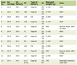 Click to view | Table 1. Comparative Performance, Strengths, Limitations, and Current Clinical Role of Diagnostic Modalities in Barrett’s Esophagus and Early Esophageal Adenocarcinoma |
| Implementation, Accessibility, and Equity | ▴Top |
The tests reviewed here are at different stages of clinical maturity. The 2025 American Gastroenterological Association surveillance guideline made no recommendation for or against WATS3D, p53 immunohistochemistry, or TissueCypher because evidence was insufficient to establish patient-important benefit [32]. Accordingly, their current role is adjunctive and context-dependent. p53 immunohistochemistry is relatively inexpensive and can be performed in many pathology laboratories. In contrast, TissueCypher and methylated-DNA assays depend on proprietary centralized testing and local reimbursement; WATS3D requires dedicated sampling and processing; non-endoscopic devices require trained staff and a pathway for confirmatory endoscopy; and cfDNA profiling and AI systems require specialized laboratory or digital infrastructure, quality control, and regulatory oversight. Cytosponge-TFF3 was cost-effective in a United Kingdom trial-based model [23], but transferability depends on disease prevalence, device and laboratory prices, uptake, downstream endoscopy capacity, and the health-system perspective.
Generalizability also remains uncertain. Many validation cohorts were recruited in Western tertiary centers and were predominantly male and non-Hispanic White; for example, the prospective EsoCheck/EsoGuard study included case and control groups that were 92% and 88% White, respectively [25]. In an ethnically diverse United States claims cohort of 12,693 patients with BE, progression estimates did not differ significantly between White and non-White groups, but only 75 esophageal cancers occurred. Lower household net worth was also associated with prevalent cancer, indicating that socioeconomic access may affect detection [35]. Prospective validation should therefore prespecify performance by sex, race and ethnicity, geography, Barrett’s segment length, community versus referral setting, and resource level. Without such evidence, a test that performs well in a selected cohort may widen rather than reduce disparities in early detection.
| Limitations of the Study | ▴Top |
Several limitations of this review should be acknowledged. First, it is a narrative rather than a systematic review. Although the search was broadened and updated, study selection and interpretation were purposive and performed by one author without a registered protocol, duplicate screening, or formal risk-of-bias assessment; selection and confirmation bias therefore cannot be excluded. Second, prioritizing open-access full text because of access constraints may have omitted relevant paywalled primary studies. Third, evidence for several advanced tests is dominated by retrospective cohorts, industry-associated validation studies, and trials conducted predominantly in high-income, non-Hispanic White populations. Diagnostic estimates may also vary with disease prevalence, spectrum, sample adequacy, assay version, and the threshold selected. These limitations restrict generalizability and leave comparative effectiveness, cost-effectiveness across health systems, optimal test intervals, and patient-important outcomes incompletely defined. Fourth, the field is advancing rapidly, and assay versions, marker panels, commercial availability, reimbursement, and regulatory status continue to evolve; performance figures should therefore be read as time-stamped estimates. Finally, this review emphasizes diagnostic and risk-stratification performance and does not systematically evaluate downstream therapeutic efficacy or the psychological and economic harms of false-positive and false-negative results.
| Conclusion | ▴Top |
BE is the recognized precursor used in current strategies for early detection of EAC, but periodic endoscopy and subjective dysplasia grading remain limited by sampling error and interobserver variability. Molecular studies refine rather than replace the classic metaplasia–dysplasia–carcinoma model. TP53 loss and clonal expansion can occur early in a subset of progressing lineages and may precede whole-genome doubling and oncogene amplification, but this route is not universal; epigenetic dysregulation, aneuploidy, and spatial clonal diversity provide complementary signals of risk. p53 immunohistochemistry and tissue systems pathology can add objectivity to biopsy interpretation, WATS3D can broaden spatial sampling, and non-endoscopic cell-collection devices can support screening or triage. Advanced biomarkers nevertheless remain adjuncts to high-quality endoscopy and expert pathology. Whether a risk-aligned pathway improves outcomes will depend on prospective clinical-utility evidence, implementation, affordability, and performance across diverse populations.
| Novelty of the Study | ▴Top |
This review examines cellular and genomic progression in BE alongside the clinical evidence for diagnostic testing. It relates each modality to the problem it addresses: p53 immunohistochemistry and tissue systems pathology add risk information to biopsy interpretation; WATS3D and AI-assisted endoscopy address lesion detection and sampling; non-endoscopic cell-collection devices support screening or triage; and cfDNA is being studied for monitoring. This organization separates established adjunctive uses from approaches that still require clinical-utility validation. It also examines how cost, infrastructure, and population representation affect implementation. The combined biological and clinical appraisal may help clinicians interpret current tools and identify priorities for further validation.
Acknowledgments
None to declare.
Financial Disclosure
This work received no specific grant from any funding agency in the public, commercial, or not-for-profit sectors.
Conflict of Interest
The author declares no conflict of interest.
Author Contributions
AS conceived the review, performed the literature search and synthesis, drafted and critically revised the manuscript, and approved the final version for submission.
Data Availability
Data sharing is not applicable to this article, as no new data were created or analyzed in this review of previously published literature.
AI Use Declaration
An AI-based large language model was used to assist with literature organization and language editing during manuscript preparation. All content, citations, interpretations, and conclusions were verified and are the sole responsibility of the author. AI tools are not listed as authors.
Abbreviations
AGA: American Gastroenterological Association; AI: artificial intelligence; BE: Barrett’s esophagus; CAD: computer-aided detection; CCNA1: cyclin A1; CDKN2A: cyclin-dependent kinase inhibitor 2A; cfDNA: cell-free DNA; DOI: digital object identifier; EAC: esophageal adenocarcinoma; EGFR: epidermal growth factor receptor; FFPE: formalin-fixed paraffin-embedded; HD: high-definition; HGD: high-grade dysplasia; HNF4A: hepatocyte nuclear factor 4 alpha; IHC: immunohistochemistry; KRAS: Kirsten rat sarcoma viral oncogene homolog; LGD: low-grade dysplasia; MYC: MYC proto-oncogene; NOTCH2: neurogenic locus notch homolog protein 2; NPV: negative predictive value; PMID: PubMed identifier; RCT: randomized controlled trial; RTK: receptor tyrosine kinase; SMAD4: SMAD family member 4; TFF3: trefoil factor 3; TP53: tumor protein p53; TSP-9: nine-biomarker tissue systems pathology test; VIM: vimentin; WATS3D: wide-area transepithelial sampling with three-dimensional computer-assisted analysis
| References | ▴Top |
- Morgan E, Soerjomataram I, Rumgay H, Coleman HG, Thrift AP, Vignat J, Laversanne M, et al. The global landscape of esophageal squamous cell carcinoma and esophageal adenocarcinoma incidence and mortality in 2020 and projections to 2040: new estimates from GLOBOCAN 2020. Gastroenterology. 2022;163(3):649-658.e642.
doi pubmed - Hvid-Jensen F, Pedersen L, Drewes AM, Sorensen HT, Funch-Jensen P. Incidence of adenocarcinoma among patients with Barrett's esophagus. N Engl J Med. 2011;365(15):1375-1383.
doi pubmed - Shaheen NJ, Falk GW, Iyer PG, Souza RF, Yadlapati RH, Sauer BG, Wani S. Diagnosis and management of Barrett's esophagus: an updated ACG guideline. Am J Gastroenterol. 2022;117(4):559-587.
doi pubmed - Fitzgerald RC, di Pietro M, Ragunath K, Ang Y, Kang JY, Watson P, Trudgill N, et al. British Society of Gastroenterology guidelines on the diagnosis and management of Barrett's oesophagus. Gut. 2014;63(1):7-42.
doi pubmed - Muthusamy VR, Wani S, Gyawali CP, Komanduri S, Participants CBsECC. AGA Clinical Practice Update on New Technology and Innovation for Surveillance and Screening in Barrett's Esophagus: Expert Review. Clin Gastroenterol Hepatol. 2022;20(12):2696-2706.e2691.
doi pubmed - Tan WK, di Pietro M, Fitzgerald RC. Past, present and future of Barrett's oesophagus. Eur J Surg Oncol. 2017;43(7):1148-1160.
doi pubmed - Nowicki-Osuch K, Zhuang L, Jammula S, Bleaney CW, Mahbubani KT, Devonshire G, Katz-Summercorn A, et al. Molecular phenotyping reveals the identity of Barrett's esophagus and its malignant transition. Science. 2021;373(6556):760-767.
doi pubmed - Ross-Innes CS, Becq J, Warren A, Cheetham RK, Northen H, O'Donovan M, Malhotra S, et al. Whole-genome sequencing provides new insights into the clonal architecture of Barrett's esophagus and esophageal adenocarcinoma. Nat Genet. 2015;47(9):1038-1046.
doi pubmed - Stachler MD, Taylor-Weiner A, Peng S, McKenna A, Agoston AT, Odze RD, Davison JM, et al. Paired exome analysis of Barrett's esophagus and adenocarcinoma. Nat Genet. 2015;47(9):1047-1055.
doi pubmed - Barchi A, Dell'Anna G, Massimino L, Mandarino FV, Vespa E, Viale E, Passaretti S, et al. Unraveling the pathogenesis of Barrett's esophagus and esophageal adenocarcinoma: the "omics" era. Front Oncol. 2024;14:1458138.
doi pubmed - Testa U, Castelli G, Pelosi E. Esophageal cancer: genomic and molecular characterization, stem cell compartment and clonal evolution. Medicines (Basel). 2017;4(3):67.
doi pubmed - Stachler MD, Bass AJ. Can genomic sequencing identify high-risk Barrett's esophagus earlier than pathologists? Cancer Cell. 2020;38(5):626-628.
doi pubmed - Ten Kate FJC, Nieboer D, Ten Kate FJW, Doukas M, Bruno MJ, Spaander MCW, Looijenga LHJ, et al. Improved progression prediction in Barrett's esophagus with low-grade dysplasia using specific histologic criteria. Am J Surg Pathol. 2018;42(7):918-926.
doi pubmed - Snyder P, Dunbar K, Cipher DJ, Souza RF, Spechler SJ, Konda VJA. Aberrant p53 immunostaining in Barrett's esophagus predicts neoplastic progression: systematic review and meta-analyses. Dig Dis Sci. 2019;64(5):1089-1097.
doi pubmed - Rajendra S, Sharma P. Barrett's esophagus. Curr Treat Options Gastroenterol. 2014;12(2):169-182.
doi pubmed - Frei NF, Khoshiwal AM, Konte K, Bossart EA, Stebbins K, Zhang Y, Pouw RE, et al. Tissue systems pathology test objectively risk stratifies Barrett's esophagus patients with low-grade dysplasia. Am J Gastroenterol. 2021;116(4):675-682.
doi pubmed - Iyer PG, Codipilly DC, Chandar AK, Agarwal S, Wang KK, Leggett CL, Latuche LR, et al. Prediction of progression in barrett's esophagus using a tissue systems pathology test: a pooled analysis of international multicenter studies. Clin Gastroenterol Hepatol. 2022;20(12):2772-2779.e2778.
doi pubmed - Davison JM, Goldblum JR, Duits LC, Khoshiwal AM, Bergman JJ, Falk GW, Diehl DL, et al. A tissue systems pathology test outperforms the standard-of-care variables in predicting progression in patients with Barrett's esophagus. Clin Transl Gastroenterol. 2023;14(11):e00631.
doi pubmed - Khoshiwal AM, Frei NF, Pouw RE, TissueCypher SLGDSPC, Smolko C, Arora M, Siegel JJ, et al. The tissue systems pathology test outperforms pathology review in risk stratifying patients with low-grade dysplasia. Gastroenterology. 2023;165(5):1168-1179.e1166.
doi pubmed - Codipilly DC, Krishna Chandar A, Wang KK, Katzka DA, Goldblum JR, Thota PN, Falk GW, et al. Wide-area transepithelial sampling for dysplasia detection in Barrett's esophagus: a systematic review and meta-analysis. Gastrointest Endosc. 2022;95(1):51-59.e57.
doi pubmed - Kaul V, Gross S, Corbett FS, Malik Z, Smith MS, Tofani C, Infantolino A. Clinical utility of wide-area transepithelial sampling with three-dimensional computer-assisted analysis (WATS3D) in identifying Barrett's esophagus and associated neoplasia. Dis Esophagus. 2020;33(12):doaa069.
doi pubmed - Fitzgerald RC, di Pietro M, O'Donovan M, Maroni R, Muldrew B, Debiram-Beecham I, Gehrung M, et al. Cytosponge-trefoil factor 3 versus usual care to identify Barrett's oesophagus in a primary care setting: a multicentre, pragmatic, randomised controlled trial. Lancet. 2020;396(10247):333-344.
doi pubmed - Swart N, Maroni R, Muldrew B, Sasieni P, Fitzgerald RC, Morris S, Consortium B. Economic evaluation of Cytosponge(R)-trefoil factor 3 for Barrett esophagus: a cost-utility analysis of randomised controlled trial data. EClinicalMedicine. 2021;37:100969.
doi pubmed - Moinova HR, LaFramboise T, Lutterbaugh JD, Chandar AK, Dumot J, Faulx A, Brock W, et al. Identifying DNA methylation biomarkers for non-endoscopic detection of Barrett's esophagus. Sci Transl Med. 2018;10(424):eaao5848.
doi pubmed - Moinova HR, Verma S, Dumot J, Faulx A, Iyer PG, Canto MI, Wang JS, et al. Multicenter, prospective trial of nonendoscopic biomarker-driven detection of Barrett's esophagus and esophageal adenocarcinoma. Am J Gastroenterol. 2024;119(11):2206-2214.
doi pubmed - Schoofs K, Ferro Dos Santos MR, De Wilde J, Roelandt S, Van de Velde S, Decruyenaere P, Meuris L, et al. Therapy response monitoring in blood plasma from esophageal adenocarcinoma patients using cell-free DNA methylation profiling. Sci Rep. 2024;14(1):31112.
doi pubmed - de Groof AJ, Struyvenberg MR, van der Putten J, van der Sommen F, Fockens KN, Curvers WL, Zinger S, et al. Deep-learning system detects neoplasia in patients with Barrett's esophagus with higher accuracy than endoscopists in a multistep training and validation study with benchmarking. Gastroenterology. 2020;158(4):915-929.e914.
doi pubmed - Novotna K, Trkova M, Pazdro A, Smejkal M, Soukupova A, Kodetova D, Smejkal P, et al. TP53 gene mutations are rare in nondysplastic Barrett's esophagus. Dig Dis Sci. 2006;51(1):110-113.
doi pubmed - Paulson TG, Galipeau PC, Oman KM, Sanchez CA, Kuhner MK, Smith LP, Hadi K, et al. Somatic whole genome dynamics of precancer in Barrett's esophagus reveals features associated with disease progression. Nat Commun. 2022;13(1):2300.
doi pubmed - Nieto T, Tomlinson CL, Dretzke J, Bayliss S, Price MJ, Dilworth M, Beggs AD, et al. A systematic review of epigenetic biomarkers in progression from non-dysplastic Barrett's oesophagus to oesophageal adenocarcinoma. BMJ Open. 2018;8(6):e020427.
doi pubmed - Jin Z, Cheng Y, Gu W, Zheng Y, Sato F, Mori Y, Olaru AV, et al. A multicenter, double-blinded validation study of methylation biomarkers for progression prediction in Barrett's esophagus. Cancer Res. 2009;69(10):4112-4115.
doi pubmed - Wani S, Zhou MJ, Sawas T, Rubenstein JH, Eluri S, Leiman DA, Sultan S, et al. AGA clinical practice guideline on surveillance of Barrett's esophagus. Gastroenterology. 2025;169(6):1184-1231.
doi pubmed - Houghton CC, Ditah I, Leggett CL, Kamboj AK, Putnam L, Sokol-Borrelli SL, Lipham JC. The tissue systems pathology test predicts risk of progression in patients with Barrett's esophagus: systematic review and meta-analysis. J Clin Gastroenterol. 2026;60(4):299-308.
doi pubmed - Kakar MT, Ramani AK, Kumar A, Lal A, Lohana DK, Radhika FNU, Mushtaq A, et al. Diagnostic test accuracy of cytosponge-trefoil factor 3 for barrett esophagus: a systemic review and meta analysis. JGH Open. 2025;9(8):e70248.
doi pubmed - Zhou MJ, Ladabaum U, Triadafilopoulos G, Clarke JO. Impact of race/ethnicity and socioeconomic status on incident and prevalent esophageal cancer in patients with Barrett's esophagus. Dis Esophagus. 2023;36(12).
doi pubmed
This article is distributed under the terms of the Creative Commons Attribution 4.0 International License (CC BY 4.0), which permits unrestricted use, distribution, and reproduction in any medium, including commercial use, provided the original work is properly cited.
Cellular and Molecular Medicine Research is published by Elmer Press Inc.