| Cellular and Molecular Medicine Research, ISSN 2817-6359 online, Open Access |
| Article copyright, the authors; Journal compilation copyright, Cell Mol Med Res and Elmer Press Inc |
| Journal website https://cmmr.elmerpub.com |
Original Article
Volume 4, Number 1, September 2026, pages 10-19

Antinuclear Antibody Seropositivity and Its Association With Transforming Growth Factor β1 and the Dynamics of Tumor-Associated Antigens in Prostate Cancer
Awatef Ben Jemaaa, b, e, Mouna Ben Azaiezc, Imen Sbeic, Sataa Sallamid, Yassine Nouirad, Ezzeddine Ghazouanic, Ridha Oueslatia
aUnit IMEC-Immunology Microbiology Environmental and Carcinogenesis, Faculty of Science of Bizerte, University of Carthage, Bizerte, Tunisia
bDepartment of Biology, Faculty of Science of Gafsa, University of Gafsa, Gafsa, Tunisia
cDepartment of Immunology, Military Hospital of Tunis, Tunis, Tunisia
dDepartment of Urology, La Rabta Hospital, University Tunis Al Manar, Tunis, Tunisia
eCorresponding Author: Awatef Ben Jemaa, Unit IMEC-Immunology Microbiology Environmental and Carcinogenesis, Faculty of Science of Bizerte, University of Carthage, Bizerte, Tunisia
Manuscript submitted July 25, 2026, accepted August 19, 2026, published online September 2, 2026
Short title: Antinuclear Antibody Seropositivity in Prostate Cancer
doi: https://doi.org/10.14740/cmmr117
| Abstract | ▴Top |
Background: The clinical significance of antinuclear antibodies (ANAs) in prostate cancer (PCa) remains unclear. This study aimed to determine the prevalence and potential clinical implications of ANA positivity in PCa patients and its association with transforming growth factor β1 (TGF-β1) and tumor-associated antigens.
Methods: ANAs were detected by indirect immunofluorescence in serum samples from 26 PCa patients. Serum prostate-specific antigen (PSA) was measured using an Immulite autoanalyzer. Immunohistochemistry was performed to assess PSA, prostate-specific membrane antigen (PSMA), CD34, and Ki-67 expression in PCa tissues. Serum TGF-β1 levels were quantified by enzyme-linked immunosorbent assay (ELISA).
Results: ANA positivity was observed in 30.8% (8/26) of PCa patients. ANA-positive and ANA-negative groups were similar in age. The most common ANA pattern was mixed, while unique cytoplasmic, speckled, cytoskeletal, and rods-and-rings patterns appeared in individual cases. ANA positivity was more frequent in patients with high Gleason scores (62.5% vs. 37.5%; P = 0.016) and high PSA levels (> 100 ng/mL). Median serum PSA was significantly higher in ANA-positive than ANA-negative patients (176 ng/mL vs. 51 ng/mL; P = 0.0046). Immunostaining for PSA, PSMA, and CD34 was significantly stronger in ANA-positive tissues (P = 0.035, P = 0.035, and P = 0.033, respectively), while Ki-67 expression showed no difference. Conversely, serum TGF-β1 levels were higher in ANA-negative patients (16.59 ng/mL vs. 11.41 ng/mL; P = 0.04).
Conclusions: Our preliminary findings suggest that ANA positivity in PCa may be associated with higher PSA levels, increased tumor antigen expression, lower serum TGF-β1 levels, and more aggressive disease features. These observations raise the possibility that ANA positivity may be associated with markers of PCa aggressiveness; however, these findings should be considered exploratory and require confirmation in larger, independent cohorts before any conclusions can be drawn regarding the potential role of ANA as a biomarker of aggressive PCa.
Keywords: Antinuclear antibodies; PSA; PSMA; TGF-β1; Prostate cancer
| Introduction | ▴Top |
Prostate cancer (PCa) is one of the leading causes of cancer-related death worldwide after lung cancer. It is also the most commonly diagnosed cancer among men aged 65 years or older, and mortality is mainly due to its late diagnosis and consequent poor prognosis [1]. Hormone therapy is used during the early stage of PCa; however, neoplastic cells no longer respond to this treatment [2]. Currently, treatments for advanced PCa have changed dramatically with the clinical development of immunotherapy [3]. Within immunotherapy strategy, the administration of antinuclear antibodies (ANAs) remains an attractive option for PCa therapy [4]. ANAs are a group of autoantibodies directed against nuclear components of cells. While ANA positivity is typically associated with autoimmune diseases such as systemic lupus erythematosus (SLE), their presence has also been documented in various malignancies, including PCa [5, 6]. Circulating ANAs in PCa reflect the complex interaction between the immune system and cancer progression. Whereas the role of cellular immunity has been investigated widely and utilized in the clinic, the importance of ANA as part of humoral immunity in antitumor activity is no longer emerging area of research [7]. Autoantibodies found in a PCa patient may be classified into two broad categories: autoantibodies against antigens that are not directly related to cancer such as ANA and autoantibodies against specific tumor antigens (tumor-associated antigens (TAAs)). In this context, TAAs such as prostate-specific antigen (PSA) and prostate-specific membrane antigen (PSMA) can elicit autoantibody responses [4, 8]. PSA is a protein produced by both normal and cancerous cells in the prostate gland. Elevated PSA levels can be an indicator of PCa condition. However, PSMA is a protein primarily found on the surface of PCa cells. While present in normal prostate tissue, PSMA expression is significantly higher in PCa cells, especially in advanced and metastatic disease [9, 10]. Moreover, PSMA imaging is valuable in identifying recurrence after surgery or radiation therapy [11]. In addition to PSA and PSMA, PCa cells highly express transforming growth factor (TGF)-β, which elicits signaling pathways to induce migration and cell metastasis [12]. As reported, the involvement of TGF-β1 in PCa is controversial. While it generally acts as tumor suppressor in normal and early-stage PCa, it could also promote tumor growth and metastasis in advanced stages of PCa disease [13, 14].
There is a lack of data on ANA in PCa, and its clinical value in the malignant prostate is not fully understood. To address this gap, we designed a study to explore the prevalence of ANA in PCa patients, to investigate its immunofluorescence pattern and to associate ANA presence with tumor characteristics. Further, we focused on evaluating ANA-positive and ANA-negative profiles by assessing the association of ANA prevalence, TGF-β1 and PCa biomarkers in PCa patients.
| Materials and Methods | ▴Top |
Chemicals and kits
Human epithelial type 2 (HEp-2) indirect immunofluorescence (IIF) was performed using commercially available HEp-2 cell substrate slides (ANA HEp-2 kit, Euroimmun, Lubeck, Germany). PSA DPC Immulite assays (Diagnostics Products Corporation, Los Angeles, CA) were used to determine serum PSA levels. The NovoLink™ Polymer Detection System (Novocastra Laboratories Ltd, Newcastle, UK) was used for immunohistochemistry analysis. The concentrations of TGF-β1 were measured by enzyme-linked immunosorbent assay (ELISA) assay (CSB-E04725h) following the manufacturer’s instructions (CUSABIO, Houston, USA). The primary antibodies used were: mouse anti-human PSMA (3E6), mouse anti-human PSA (ER-PR8), mouse anti-human CD34 (QBend10) (Dako, Glostrup, Denmark) and mouse anti-human Ki-67 (CUSABIO, Houston, USA).
Study design and patients
This was a retrospective study of patients with malignant prostate undergoing radical prostatectomy at La Rabta Hospital (Tunis, Tunisia). Following approval by the Institute Ethical Committee (6/2022) and written informed consent from all the participants in accordance with the Declaration of Helsinki, we retrospectively recruited 26 patients with PCa aged 58 to 85 years in the study. Baseline laboratory and clinical data including demographic details of the limited number of cases were retrieved from a medical record system. All pathological, clinical, and personal data were anonymized and separated from personal identifiers.
Sampling for serology assays
Blood samples were processed according to hospital’s standard procedures. The peripheral blood samples were strictly collected for each study subject. For the serological testing, 5 mL of whole blood was collected in a tube without anticoagulant. The serum was allowed to separate (centrifuged for 20 min at 2,000 rpm), aliquoted, and kept at −80 °C until ANA, PSA and TGF-β1 tests were performed.
Sampling for immunohistochemistry analysis
After radical prostatectomy, prostate tissues were fixed at room temperature in 0.1 M phosphate-buffered 10% formaldehyde, dehydrated and embedded in paraffin. Sections (3 µm thick) were processed. Thereafter, the primary antibodies (PSA, PSMA, CD34 and Ki-67) were detected by immunohistochemistry analysis.
Indirect immunofluorescence on HEp-2 cells
All the samples were screened to ANA by indirect immunofluorescence on HEp-2 cells, using the commercially available kit ANA HEp-2 (Euroimmun, Lubeck, Germany) according to the manufacturer’s instructions. The fluorescence patterns were interpreted as cytoplasmic, speckled, cytoskeletal, membranous, rods and rings. A titer of 1:160 or higher was considered to indicate ANA positivity. This cutoff was selected because low ANA titers, particularly 1:40 and 1:80, may occur relatively frequently in healthy individuals and may therefore have limited specificity. In contrast, a titer of 1:160 provides a more conservative threshold that reduces the likelihood of considering low-level, nonspecific ANA reactivity as clinically relevant. A previous study has reported that serum ANA cutoff of 1:160 serum dilution has high specificity with the ability to exclude 95% of the normal samples [15], and thus we classified serum ANA cutoff at 1:160 and divided PCa patients into the ANA positive (ANA ≥ 1:160) and ANA-negative (ANA < 1:160) groups. The clinical and histopathological characteristics of patients with PCa were compared between the ANA-positive and ANA-negative groups.
PSA immunoassays
PSA DPC Immulite assays (Diagnostics Products Corporation, Los Angeles, CA) were used to determine serum PSA levels in these patients before radical prostatectomy. These assays were performed according to the manufacturer’s instructions. This was a double-site sandwich immunoassay performed in the solid phase, using the monoclonal and polyclonal anti-PSA antibodies. The reaction was developed with an enhanced chemiluminescence. The normal PSA level by DPC was 4 ng/mL.
TGF-β1 immunoassays
TGF-β1 was assessed in PCa serum samples. The concentrations of TGF-β1 were measured by ELISA assay (CSB-E04725h) following the manufacturer’s instructions (CUSABIO, Houston, USA). It is a sandwich immunoassay. Quantification of serum TGF-β1 levels were performed before radical prostatectomy.
Immunohistochemistry analysis
Immunohistochemistry analysis was processed following the NovoLink™ Polymer Detection Systems (Novocastra Laboratories Ltd, Newcastle, UK) method. The primary antibodies used were: mouse anti-human PSMA (3E6), mouse anti-human PSA (ER-PR8), mouse anti-human CD34 (QBend10) (Dako, Glostrup, Denmark) and mouse anti-human Ki-67 (CUSABIO, Houston, USA). CD34 antibody was used to label vessels in the prostate tissues.
Sections (3 µm thick) were deparaffinized, rehydrated through graded alcohols and washed in deionized water. To retrieve antigens, sections were incubated in citric acid solution (0.1 M, pH 6) for 20 min in 98 °C using a water bath. Slides were allowed to cool for another 20 min, followed by washing in deionized water. Endogenous peroxidase activity was quenched by incubation with peroxidase block for 5 min. Each incubation step was carried out at room temperature and was followed by two sequential washes (5 min each) in TBS. Sections were incubated with protein block for 5 min to prevent nonspecific binding of the first antibody. Thereafter, the primary antibodies were applied at a dilution of 1:50 (PSMA), 1:100 (PSA, CD34, Ki-67) in antibody diluents (Dako, Glostrup, Denmark) at room temperature for 30 min. Afterwards, the sections were incubated with post primary block for 30 min to block nonspecific polymer binding. The sections were incubated with NovoLink™ Polymer for 30 min followed by incubations with 3,3’-diaminobenzidine (DAB) working solution for 5 min to develop peroxidase activity. Slides were counterstained with hematoxylin and mounted. Staining specificity was checked using negative controls. Prostatic tissues of each type were incubated in blocking peptides (Santa Cruz Biotechnology, Santa Cruz, CA, USA) instead of primary antibodies.
The observer performing the measurements was blinded to the experimental group and sample identity to minimize potential observer bias. In addition, the measurements were repeated on a randomly selected subset of sections to assess intra-observer reproducibility. The repeated measurements showed good agreement, confirming the reproducibility of the quantitative assessment.
Of each prostate, six histological sections were selected at random. In each section, the staining intensity (optical density (OD)) per unit surface area was measured with an automatic image analyzer (Motic Images Advanced version 3.2, Motic China Group Co., China) in five light microscopic fields per section, using the × 40 objective. The intensity of immunohistochemical staining was quantified by measuring the OD of the positive staining within a defined tissue surface area. OD was expressed relative to the analyzed surface area in order to account for differences in the size of the regions evaluated and to provide a standardized measure of staining intensity. The regions of interest corresponding to the immunopositive areas were manually delineated by the investigator using the computer mouse and the image-analysis software. Care was taken to accurately delimit the tissue areas to be analyzed while excluding background regions, tissue artifacts, folds, tears, and other non-representative areas. The image analyzer then automatically calculated the OD within the manually selected areas. Delimitation of surface areas was carried out manually using the mouse of the image analyzer. To correct for nonspecific background staining and intrinsic OD of the tissue, a negative control section was analyzed for each immunostained section. For each positive immunostained section, one negative control section (the following in a series of consecutive sections) was also used, and the optic density of this control section was taken away from that of the stained section.
Statistical analysis
All statistical analyses were performed by GraphPad Prism software 5 (GraphPad PRISMA 5.0 computer program). Shapiro–Wilk normality test was conducted to estimate the distribution of the data. Categorical variables were expressed as number or percentages, and significance was detected by Fisher’s exact test. Continuous variables were expressed as mean and range or median and interquartile range (IQR) values. For normally distributed continuous variables, differences between groups were compared using Unpaired t-test; conversely, the Mann–Whitney U-test was used for continuous variables that were not normally distributed. For all statistical analysis, P < 0.05 was considered statistically significant.
| Results | ▴Top |
Comparison of PCa patient characteristics depending on the ANA status
A total of 26 patients suffered from PCa with median age (range) 74 years (58–85) were enrolled in this study. Patients were categorized into two groups: ANA-positive (ANA ≥ 1:160) and ANA-negative (ANA < 1:160). Comparative evaluations of PCa patients with and without ANA have been detailed in Table 1. ANAs were found in 30.8% (8/26) of the PCa patients. The proportion of patients with ANA-negative was 69.2% (18/26) of the PCa patients. The ANA-positive group had a similar median age (range) (75 years (68–83)) as compared to ANA-negative group (75 years (58–85), P = 0.977). The patients with ANA-positive status had a lesser frequency among patients with Gleason score 6–7 as compared to patients with Gleason score 8–10 (37.5% vs 62.5%; P = 0.016) (Table 1). Moreover, the ANA-positive group had a significantly higher median (IQR) sera PSA level (176 ng/mL (83.5–1203) vs 51 ng/mL (22.98–100); P = 0.0046)) as compared to ANA-negative group (Table 1). Half of PCa patients with positive ANA received hormone treatment. Likewise, 55.6% (10/18) of patients received hormone treatment in ANA-negative group (Table 1). There was no significant difference in response to hormone therapy between ANA-positive and ANA-negative groups (P > 0.05). In the ANA-positive group, 25% (2/8) of patients did not respond to hormone therapy after a mean time of 12 months of treatment. Similarly, in the ANA-negative group, few patients did not respond to hormonal treatment (11.1% (2/18)). Furthermore, there was no significant difference between these two groups concerning metastasis of PCa (P > 0.05). Only one PCa patient developed metastasis in either the ANA-positive or ANA-negative groups (Table 1).
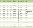 Click to view | Table 1. Comparison of Malignant Prostate Lesions Characteristics According to Positivity of ANA (Positive ANA PCa Patients N = 8; Negative ANA PCa Patients N = 18) |
Details of the ANA patterns have been described in Table 1 and Figure 1. Immunofluorescence showed a diversity of autoantibody patterns with cytoplasmic, speckled, cytoskeletal, rods and rings in one PCa patient for each ANA pattern. Nevertheless, the most common pattern was mixed pattern (50% (4/8) of ANA-positive patients). Mixed patterns of ANA, including speckled, midbody, nucleolar, and other cytoplasmic fluorescence, interfered with their detection (Table 1, Fig. 1).
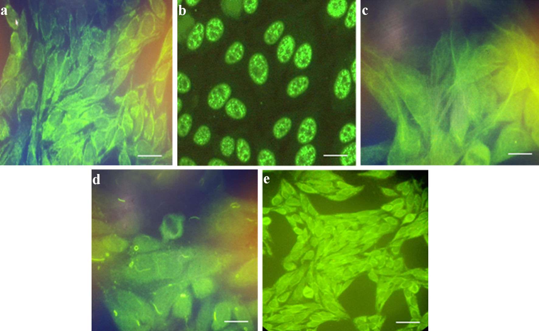 Click for large image | Figure 1. Representative images of ANA patterns in positive sera from patients with malignant prostate disease (n = 8): (a) cytoplasmic; (b) speckled; (c) cytoskeletal; (d) rods and rings; and (e) mixed pattern. Mixed patterns of ANA included speckled, midbody, nucleolar, and other cytoplasmic fluorescence, which interfered with their detection. Scale bar = 100 µm. ANA: antinuclear antibody. |
Immunoreactivity to PCa biomarkers (PSA and PSMA) in ANA-positive and ANA-negative groups
To understand the association between PCa biomarkers and ANA status, we used immunohistochemistry analysis to evaluate the expression of PSA and PSMA in PCa tissues. As shown in Figure 2, PSA exhibited low cytoplasmic staining; whereas PSMA was overexpressed in malignant prostate tissues. Furthermore, PSMA has characteristic cytoplasmic but also strong membranous staining (Fig. 2). Notably, our investigation based on ANA status revealed a significant higher immunostaining to PSA in ANA-positive as compared to ANA-negative group (15.3 ± 3.3 vs 4.39 ± 2.37; P = 0.035) (Fig. 3). Likewise, immunoreactivity to PSMA was significantly more elevated in ANA-positive as compared to ANA-negative group (45.3 ± 3.22 vs 24.71 ± 3.71; P = 0.035) (Fig. 3).
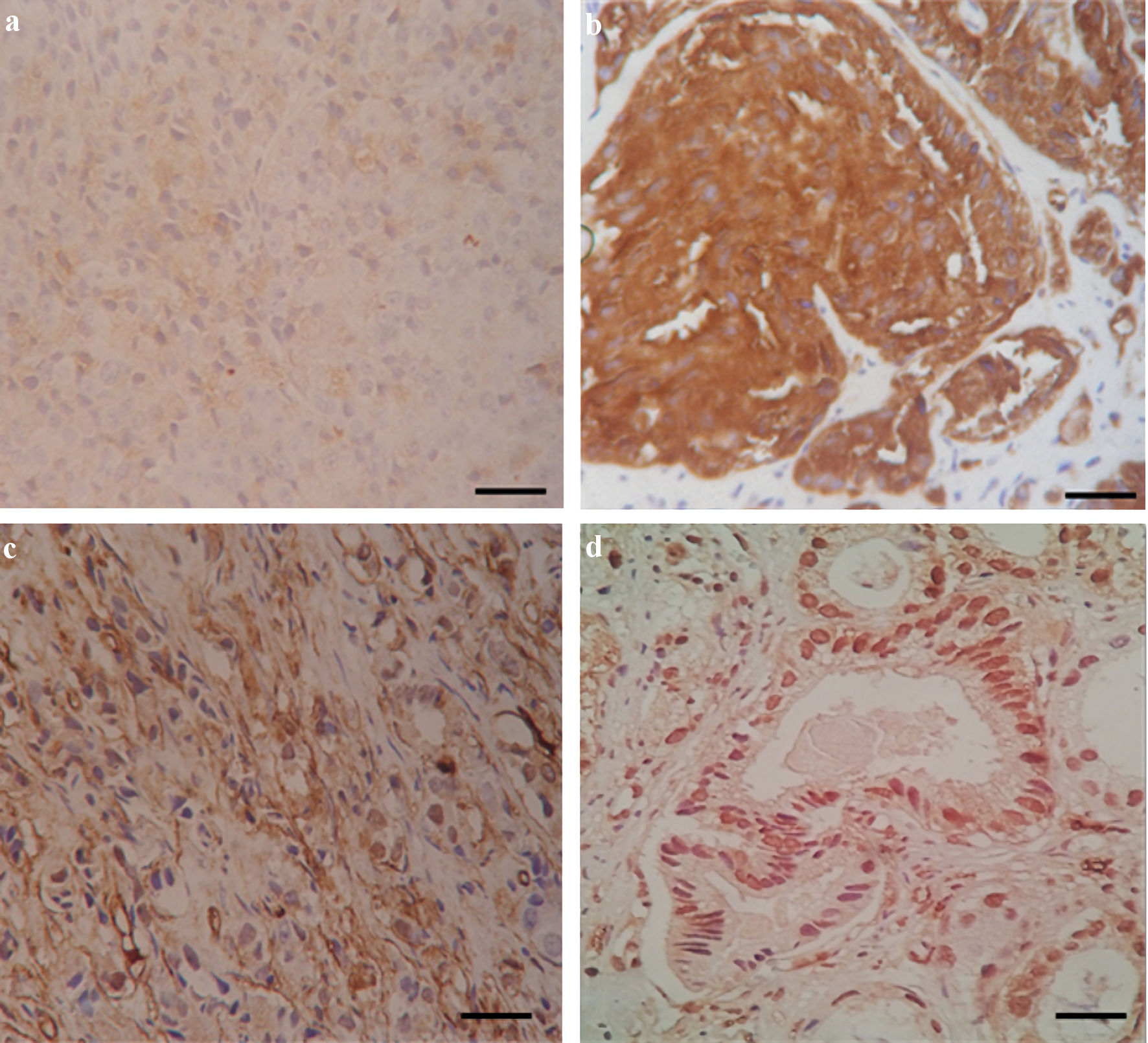 Click for large image | Figure 2. Immunostaining of PSA, PSMA, CD34, and Ki-67 expression in human prostate cancer tissues (n = 26). (a) PSA shows low cytoplasmic staining. (b) PSMA has characteristic cytoplasmic but also strong membranous staining. (c) CD34 immunoreactivity is restricted to the blood vessels. (d) Ki-67 shows high nuclear staining. Scale bar = 100 µm. PSA: prostate-specific antigen; PSMA: prostate-specific membrane antigen. |
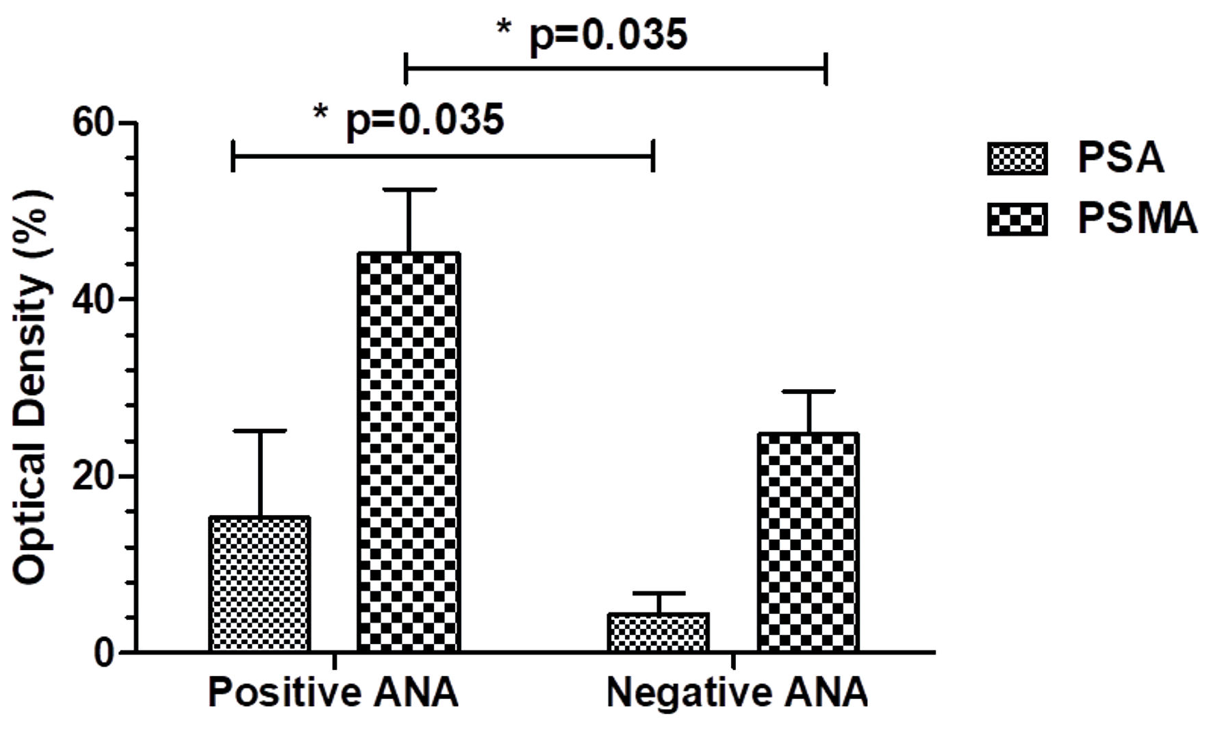 Click for large image | Figure 3. Comparison of PSA and PSMA immunoreactivity in malignant prostate patients depending on the ANA positivity (positive ANA PCa patients n = 8; negative ANA PCa patients n = 18). *P < 0.05. ANA: antinuclear antibody; PSA: prostate-specific antigen; PCa: prostate cancer; PSMA: prostate-specific membrane antigen. |
Proliferation index and angiogenesis depending on the ANA status in malignant prostate
To further evaluate the role of ANA in PCa progression, we performed immunohistochemistry assay to detect the proliferation index Ki-67 in malignant prostate tissues. As shown in Figure 2, a strong immunoreactivity to Ki-67 was restricted to the nuclear of prostate tumor cells. However, our finding showed no significant variation in Ki-67 expression between ANA-positive and ANA-negative groups (23.78 ± 1.13 vs 25.321 ± 1.83; P = 0.31) (Fig. 4). Next, the study assessed the association between ANA status and angiogenesis in PCa tissues. CD34 was used as biomarker of angiogenesis in malignant prostate tissues. Our results found that strong CD34 immunoreactivity was restricted to the blood vessels of PCa tissues (Fig. 2). Interestingly, CD34 exhibited higher immunostaining in ANA-positive group as compared to ANA-negative group (17.02 ± 1.74 vs 9.99 ± 1.06; P = 0.033) (Fig. 4).
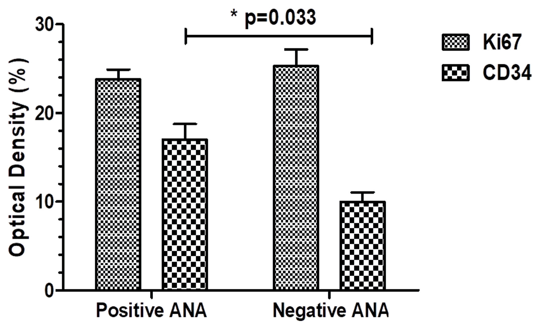 Click for large image | Figure 4. Ki67 and CD34 immunoexpression among prostate cancer patients according to the ANA positivity (positive ANA PCa patients n = 8; negative ANA PCa patients n = 18). *P < 0.05. ANA: antinuclear antibody; PCa: prostate cancer. |
Comparison of serum TGF-β1 levels in ANA-positive and ANA-negative PCa groups
To further investigate whether the expression of TGF-β1 is influenced by ANA status in PCa, we performed ELISA assay to evaluate TGF-β1 expression in sera of malignant prostate patients. As shown in Figure 5, we revealed significantly elevated TGF-β1 levels in ANA-negative group as compared to ANA-positive group (16.59 ± 1.59 ng/ml vs 11.41 ± 2.47 ng/mL; P = 0.04) (Fig. 5).
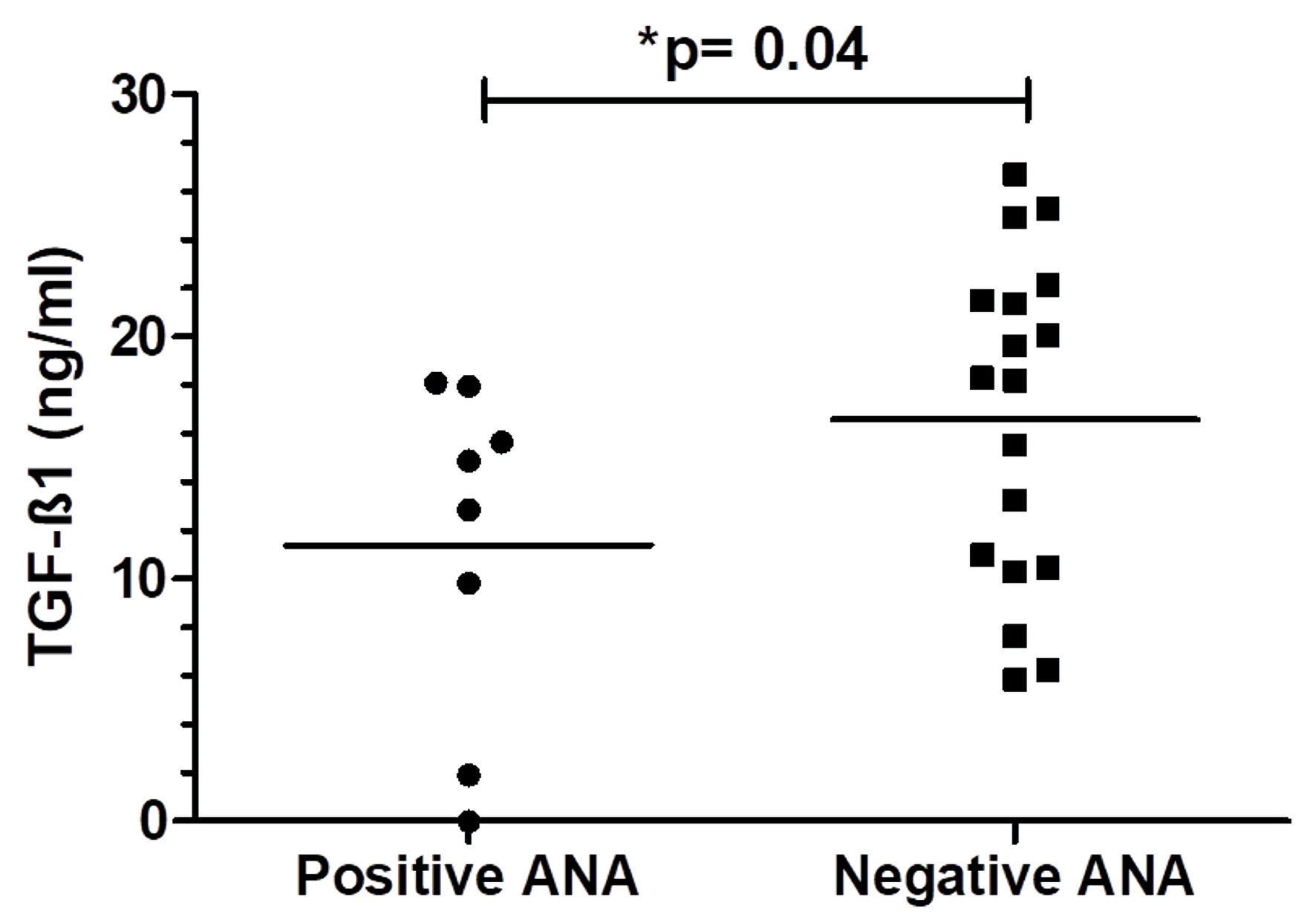 Click for large image | Figure 5. TGF-β1 levels among prostate cancer patients depending on the ANA positivity (positive ANA PCa patients n = 8; negative ANA PCa patients n = 18). *P < 0.05. ANA: antinuclear antibody; TGF-β1: transforming growth factor β1; PCa: prostate cancer. |
| Discussion | ▴Top |
Our results showed that PCa patients have a scanty prevalence of positivity for ANA. These findings agree with those of Mohammad et al [6], who studied 12 newly diagnosed PCa patients and found that only five cases of PCa patients were ANA positive. According to Sakakida et al [16], positive ANA titers were shown in nine out of 191 patients with non-small cell lung cancer. Another notable finding of the present study was that ANA positivity appeared to be associated with several PCa characteristics, including Gleason score and serum PSA levels. Our finding showed that the ANA-positive group had significantly higher sera PSA compared to the ANA-negative group. Moreover, ANA positivity appeared to be more frequent among PCa patients with a high Gleason score than among those with an intermediate Gleason score. Previous studies have reported that ANA was frequently found in the sera of patients with solid neoplasms of the breast, colon, and lungs, or with lymphoma [17–19]. Additionally, the prevalence of ANA positivity was higher in patients with breast cancer than in patients with benign lesions and controls [20]. An overrepresentation of ANA was confirmed and persisted in the cancer group with malignant pelvic disease compared to the benign pelvic disease group [21]. In the case of chronic liver disease, patients who developed hepatocellular carcinoma showed that the prevalence of patients with positive ANA increased from 27% of cases prior to cancer diagnosis to 40% just before the cancer appearance. Furthermore, in those who were negative, nearly 30% of them converted to positive ANA when the cancer was detected [22]. Based on the results, ANA could play roles in disease aggressiveness and promoting PCa cell proliferation.
Several studies reported the existence of growing evidence of a bidirectional relationship between an immune response and cancer through autoantibodies. These findings showed that the immune system of such patients reacts to factors involved in carcinogenesis and that ANA, as part of this response, may not only be used as biomarkers of cancer and disease outcome, but also to characterize the tumor immune response. Autoantibodies found in cancer subjects could be attributed to several cellular and humoral immunological aberrations, which occur during the tumorigenesis process [23, 24]. Therefore, the observed presence of autoantibodies in PCa may reflect, at least in part, the complex interplay between immune responses and tumor progression. ANA belonged to the category of autoantibodies that reacted to antigens that were not directly associated with the tumor. ANA production may represent a consequence of tumor-associated inflammation, tissue damage, oxidative stress, or increased exposure to TAAs rather than a direct contributor to tumor progression. These autoantibodies found in cancer patients could be involved in the regulation of cell cycle and mitosis [25]. The detection of ANA is traditionally performed with immunofluorescent techniques using human epithelial cell lines (HEp-2) as substrates. These techniques unravel distinct staining patterns that are highly valued in the tumorigenesis process. Staining patterns that act as fingerprints, such as homogeneous, nucleolar, speckled, and cytoplasmic patterns, were indicative of potential connections to cancer aggressiveness [26, 27]. In this study, it was found that numerous ANA patterns were present in PCa patients, such as cytoplasmic, speckled, cytoskeletal, rods and rings patterns. However, a mixed pattern of ANA was the most frequently observed in the current study. Studies have shown that a nucleolar ANA pattern was associated with a higher risk of cancer, including leukemia, and may indicate a more aggressive form of the disease, while homogeneous and speckled ANA patterns were often linked to a good cancer prognosis [20, 26, 28]. A mixed pattern should not be ignored and could be indicative of certain tumors [29].
As with other cancer types, in PCa, TAAs such as PSA and PSMA could elicit autoantibody responses. Consequently, the immune system might recognize abnormal expression or post-translational modifications of these proteins as foreign, triggering an autoimmune response [4, 8]. Depending on ANA positivity, it was found that both PSA and PSMA were significantly more highly expressed in the ANA-positive group compared to the ANA-negative group. These findings suggested an association between upregulation of PCa biomarkers (PSA and PSMA) and ANA expression in the context of prostate malignant disease. In a prior study, we found that PCa patients with the (PSA+, PSMA+) phenotype could be indicative of advanced PCa with poor prognosis. PSMA was overexpressed in this PCa group with the (PSA+, PSMA+) phenotype [9]. Another notable finding of the present study was that CD34 expression appeared to be higher in ANA-positive PCa patients than in ANA-negative patients. In PCa, CD34 is often used as a marker to assess angiogenesis, which is essential for PCa cells to spread and develop metastasis. Moreover, overexpression of CD34 might indicate more aggressive PCa and a higher risk of recurrence after treatment [30]. According to the current results, ANA were more indicative of aggressive PCa and played a role in promoting PCa development and progression. The presence of autoantibodies could indicate that ANA blocks tumor-suppressive pathways, promoting inflammation and tumor progression of PCa. Our findings agree with those of Skare et al [31], which found that ANA was more common in invasive cervical lesions than in controls or noninvasive lesions. Several studies found significant ANA positivity in breast, colon, lungs and lymphoma cancer patients compared to benign and controls, especially in advanced cases [17–19]. Conversely, some authors agree with anti-tumor effect of ANA in patients with neoplastic disease. They suggested that the immune system of these patients may respond to factors involved in carcinogenesis, with ANA production potentially representing part of this immune response and being associated with a protective effect against tumor progression. Thus, the presence of autoantibodies may reflect an ongoing immune response and potentially contribute to immune surveillance against tumor cells [32, 33]. Experimental studies have shown that ANA could enhance antibodies-dependent cellular cytotoxicity (ADCC) and cytokine production, promoting the activity of immune system [32, 33]. This idea was supported by the fact that a monoclonal tumor cell surface-reactive ANA suppressed the growth of an aggressive PCa cells [4].
In our cohort, no significant association was observed between ANA positivity and the Ki-67 proliferation index in patients with PCa. As in other solid tumors, Ki-67 is a commonly used marker to assess the rate of PCa cell proliferation. Furthermore, Ki-67 might predict PCa aggressiveness and resistance to the treatment [34]. However, a previous study showed variability of Ki-67 in high-grade PCa, with surprisingly low Ki-67 expression in a subset of cases [35].
Accumulating evidence has shown the involvement of the multifunctional cytokine TGF-β1 in PCa progression and in immune response against tumor cells [36]. One of the novel findings of this study is that serum TGF-β1 level in the ANA-positive PCa group was lower than that in ANA-negative PCa group. Furthermore, ANA positivity was more observed in advanced cases of PCa patients with high Gleason grade. It was previously reported that serum TGF-β1 level correlated with prostate disease grade, clinical stage and metastasis [37, 38]. These findings, together with our preliminary data, suggest a possible inverse association between ANA positivity and TGF-β1 levels in the context of PCa. The present study cannot establish a causal relationship between ANA positivity and reduced TGF-β1 levels. It remains unclear whether altered TGF-β1 levels contribute to autoantibody production, are a consequence of tumor-associated immune alterations, or represent an indirect association.
The effect of TGF-β1 on PCa cell proliferation and its involvement in immune response against PCa remained controversial. Both inhibition and promoting PCa cell proliferation and aggressiveness were reported. While it generally inhibited cell growth in normal and early-stage PCa, it could also promote tumor growth and metastasis in later stages of PCa disease [13, 14]. Nevertheless, Reis et al [39] showed that decreased TGF-β expression was associated with PCa progression. Additionally, TGF-β could inhibit the activity of immune cells like T cells, natural killer (NK) cells, and dendritic cells, preventing them from effectively attacking cancer cells [40, 41].
In PCa, one of the best-characterized nuclear autoantigens is lens epithelium-derived growth factor p75 (LEDGF/p75), also known as DFS70. Daniels et al [42] demonstrated that LEDGF/p75 is a target of antinuclear autoantibodies in patients with PCa and proposed that its high expression in prostate tumors and release during apoptosis may contribute to the development of this humoral immune response. Research increasingly suggests that the ongoing production of tumor-associated autoantibodies and chronic inflammatory responses in PCa could result in the infiltration and activation of immunosuppressive cells. This process established an immunosuppressive microenvironment that promotes prostate tumor immune evasion [43].
Our study tried to overcome the shortcomings of the previous studies, but we still have some limitations including the relatively small number of paired samples, which limits the statistical power of the analysis and may restrict the generalizability of the findings. Future studies should include larger, independent, and preferably multicenter cohorts to validate the present findings and allow for more robust statistical analyses and control of the false-positive rate when multiple comparisons are performed. Second, the titers of autoantibodies directed against PSA and PSMA, were not quantitatively assessed. Future investigations should incorporate quantitative measurement of these autoantibody responses to determine their association with clinical and pathological characteristics and to evaluate their potential diagnostic or prognostic value. Importantly, without an appropriate control group, it is not possible to determine whether the observed ANA positivity is specific to PCa or may also occur in healthy individuals or patients with benign prostatic conditions. Consequently, the present findings should be interpreted as describing the association of ANA positivity with clinicopathological characteristics within the PCa cohort rather than establishing ANA as a PCa-specific biomarker. Finally, the molecular assays were evaluated in a limited manner. Further studies should incorporate a more comprehensive molecular characterization using validated and quantitative molecular assays. Such approaches could provide additional insight into the underlying molecular mechanisms and help determine whether the observed findings can be translated into clinically relevant biomarkers. Future prospective studies involving larger and well-characterized cohorts, paired samples, including patients with different stages of PCa, particularly advanced and metastatic disease, quantitative autoantibody measurements, and comprehensive molecular analyses are therefore warranted to validate and extend the findings of the present study.
Conclusions
Taken together, although ANA prevalence was relatively low among patients with PCa, ANA positivity appeared to be more frequent in patients with advanced disease and higher Gleason scores. ANA positivity was also associated with higher expression of the prostate TAAs PSA and PSMA and increased CD34 expression, whereas serum TGF-β1 levels were lower in ANA-positive than in ANA-negative patients. These findings suggest a possible association between ANA positivity and features of more advanced PCa. However, given the limited sample size and exploratory nature of this study, these results should be considered preliminary and hypothesis-generating rather than definitive evidence of clinical utility. ANA may represent a potentially promising biomarker of disease characteristics in PCa, but its diagnostic or prognostic value requires confirmation in larger, independent, and preferably prospective multicenter studies with adequate follow-up and comprehensive assessment of autoantibody profiles. Such studies may also provide further insight into the immunobiological mechanisms underlying ANA production in PCa and determine its potential relevance in clinical practice.
Acknowledgments
We thank all patients for their contribution to the study. The authors thank the medical and technical staffs of the Department of Immunology, Military Hospital of Tunis, Tunisia, and the Department of Urology, La Rabta Hospital, Tunis, Tunisia, for their precious contribution in the study.
Financial Disclosure
This study was supported in part by the Tunisian Ministry of High Education and Scientific Research from the Research Fund Program for the Encouragement of Young Researchers (21PEJC D5P14).
Conflict of Interest
The authors declare that they have no known competing financial interests or personal relationships that could have appeared to influence the work reported in this paper.
Informed Consent
Written informed consent was obtained from all participants prior to their inclusion in the study. All pathological, clinical and personal data were anonymized and separated from any personal identifiers.
Author Contributions
Awatef Ben Jemaa: investigation, conceptualization, methodology, data curation, writing – review and editing. Mouna Ben Azaiez: investigation, conception, design of the study, methodology, data curation and writing. Imen Sbei: data curation and investigation. Sataa Sallami: data curation and investigation. Yassine Nouira: data curation and investigation. Ezzeddine Ghazouani: data curation and investigation. Ridha Oueslati: conception and design of the study.
Data Availability
The datasets used and/or analyzed during the current study are available from the corresponding author on reasonable request.
Abbreviations
ANAs: antinuclear antibodies; LEDGF/p75: lens epithelium-derived growth factor p75; PCa: prostate cancer; PSA: prostate-specific antigen; PSMA: prostate-specific membrane antigen; TGF-β1: transforming growth factor β1
| References | ▴Top |
- Belkahla S, Nahvi I, Biswas S, Nahvi I, Ben Amor N. Advances and development of prostate cancer, treatment, and strategies: a systemic review. Front Cell Dev Biol. 2022;10:991330.
doi pubmed - Shiner A, Sperandio RC, Naimi M, Emmenegger U. Prostate cancer liver metastasis: an ominous metastatic site in need of distinct management strategies. J Clin Med. 2024;13(3):734.
doi pubmed - Sridaran D, Bradshaw E, DeSelm C, Pachynski R, Mahajan K, Mahajan NP. Prostate cancer immunotherapy: Improving clinical outcomes with a multi-pronged approach. Cell Rep Med. 2023;4(10):101199.
doi pubmed - Jayakrishnan R, Schafer C, Tan SH. Prostate cancer autoantibodies - applications in diagnosis, prognosis, monitoring disease progression and immunotherapy. Am J Clin Exp Urol. 2023;11(2):79-102.
pubmed - Robertson JM, James JA. Preclinical systemic lupus erythematosus. Rheum Dis Clin North Am. 2014;40(4):621-635.
doi pubmed - Mohammad DO, Najim RH. Relation of anti nuclear antibodies with different types of cancer. Tikrit J Pharma Sci. 2011;7:51-54.
- Torchilin VP, Iakoubov LZ, Estrov Z. Antinuclear autoantibodies as potential antineoplastic agents. Trends Immunol. 2001;22(8):424-427.
doi pubmed - Kiessling A, Wehner R, Fussel S, Bachmann M, Wirth MP, Schmitz M. Tumor-associated antigens for specific immunotherapy of prostate cancer. Cancers (Basel). 2012;4(1):193-217.
doi pubmed - Ben Jemaa A, Bouraoui Y, Sallami S, Nouira Y, Oueslati R. A comparison of the biological features of prostate cancer with (PSA+, PSMA+) profile according to RKIP. Biomed Res Int. 2013;2013:409179.
doi pubmed - Giraudet AL, Kryza D, Hofman M, Moreau A, Fizazi K, Flechon A, Hicks RJ, et al. PSMA targeting in metastatic castration-resistant prostate cancer: where are we and where are we going? Ther Adv Med Oncol. 2021;13:17588359211053898.
doi pubmed - Houshmand S, Lawhn-Heath C, Behr S. PSMA PET imaging in the diagnosis and management of prostate cancer. Abdom Radiol (NY). 2023;48(12):3610-3623.
doi pubmed - Bjerke GA, Yang CS, Frierson HF, Paschal BM, Wotton D. Activation of Akt signaling in prostate induces a TGFbeta-mediated restraint on cancer progression and metastasis. Oncogene. 2014;33(28):3660-3667.
doi pubmed - Kim IY, Ahn HJ, Zelner DJ, Shaw JW, Sensibar JA, Kim JH, Kato M, et al. Genetic change in transforming growth factor β (TGF-β) receptor type I gene correlates with insensitivity to TGF-β1 in human prostate cancer cells. Cancer Res. 1996;56:44-48.
- Sun DY, Wu JQ, He ZH, He MF, Sun HB. Cancer-associated fibroblast regulate proliferation and migration of prostate cancer cells through TGF-beta signaling pathway. Life Sci. 2019;235:116791.
doi pubmed - Tan EM, Feltkamp TE, Smolen JS, Butcher B, Dawkins R, Fritzler MJ, Gordon T, et al. Range of antinuclear antibodies in "healthy" individuals. Arthritis Rheum. 1997;40(9):1601-1611.
doi pubmed - Sakakida T, Ishikawa T, Chihara Y, Harita S, Uchino J, Tabuchi Y, Komori S, et al. Safety and efficacy of PD-1/PD-L1 blockade in patients with preexisting antinuclear antibodies. Clin Transl Oncol. 2020;22(6):919-927.
doi pubmed - Imran A, Neelam F, Tariq M. Incidence of circulating antinuclear antibodies in cancer patients. Indian J Med Sci. 2003;57(3):113-116.
pubmed - Solans-Laque R, Perez-Bocanegra C, Salud-Salvia A, Fonollosa-Pla V, Rodrigo MJ, Armadans L, Simeon-Aznar CP, et al. Clinical significance of antinuclear antibodies in malignant diseases: association with rheumatic and connective tissue paraneoplastic syndromes. Lupus. 2004;13(3):159-164.
doi pubmed - Zou HY, Gu X, Yu WZ, Wang Z, Jiao M. Detection of serum antinuclear antibodies in lymphoma patients. Genet Mol Res. 2015;14(4):16546-16552.
doi pubmed - Nisihara R, Machoski MCC, Neppel A, Maestri CA, Messias-Reason I, Skare TL. Anti-nuclear antibodies in patients with breast cancer. Clin Exp Immunol. 2018;193(2):178-182.
doi pubmed - Heegaard NH, West-Norager M, Tanassi JT, Houen G, Nedergaard L, Hogdall C, Hogdall E. Circulating antinuclear antibodies in patients with pelvic masses are associated with malignancy and decreased survival. PLoS One. 2012;7(2):e30997.
doi pubmed - Imai H, Nakano Y, Kiyosawa K, Tan EM. Increasing titers and changing specificities of antinuclear antibodies in patients with chronic liver disease who develop hepatocellular carcinoma. Cancer. 1993;71(1):26-35.
doi pubmed - Reuschenbach M, von Knebel Doeberitz M, Wentzensen N. A systematic review of humoral immune responses against tumor antigens. Cancer Immunol Immunother. 2009;58(10):1535-1544.
doi pubmed - Zaenker P, Gray ES, Ziman MR. Autoantibody production in cancer—the humoral immune response toward autologous antigens in cancer patients. Autoimmun Rev. 2016;15(5):477-483.
doi pubmed - Ortona E, Pierdominici M, Berstein L. Autoantibodies to estrogen receptors and their involvement in autoimmune diseases and cancer. J Steroid Biochem Mol Biol. 2014;144(Pt B):260-267.
doi pubmed - Gauderon A, Roux-Lombard P, Spoerl D. Antinuclear antibodies with a homogeneous and speckled immunofluorescence pattern are associated with lack of cancer while those with a nucleolar pattern with the presence of cancer. Front Med (Lausanne). 2020;7:165.
doi pubmed - Koga T, Okamoto M, Satoh M, Fujimoto K, Zaizen Y, Chikasue T, Sumi A, et al. Positive autoantibody is associated with malignancies in patients with idiopathic interstitial pneumonias. Biomedicines. 2022;10(10):2469.
doi pubmed - Wang R, Zhao H, Liu Y, Kang B, Cai J. Antinuclear antibodies with a nucleolar pattern are associated with a significant reduction in the overall survival of patients with leukemia: a retrospective cohort study. Front Oncol. 2021;11:631038.
doi pubmed - Cerimagic A, Dzananovic N. Antinuclear antibodies (ANA) patterns in paraneoplastic cerebellar degeneration during the course of disease and treatment protocols - A case report. J Transl Autoimmun. 2020;3:100034.
doi pubmed - Gautam KA, Singh AN, Srivastav AN, Sankhwar SN. Angiogenesis in prostate cancer and benign prostatic hyperplasia assessed by VEGF and CD-34 IHC: a comparative clinico-pathological study. African Journal of Urology. 2018;24(2):98-103.
- Skare TL, Neppel A, Machoski MCC, Maestri CA, Messias-Reason I, Nisihara R. Antinuclear antibodies in patients with cervical lesions and invasive cervical cancer. Immunol Lett. 2019;208:8-10.
doi pubmed - Kobold S, Lutkens T, Cao Y, Bokemeyer C, Atanackovic D. Autoantibodies against tumor-related antigens: incidence and biologic significance. Hum Immunol. 2010;71(7):643-651.
doi pubmed - Hock BD, Goddard L, Dobson LJ, MacPherson SA, O'Donnell JL, McKenzie JL, McLellan AD. Impact of rheumatoid factors on the function of therapeutic monoclonals specific for PD-1/PD-L1. Cancer Immunol Immunother. 2025;74(7):216.
doi pubmed - Berney DM, Gopalan A, Kudahetti S, Fisher G, Ambroisine L, Foster CS, Reuter V, et al. Ki-67 and outcome in clinically localised prostate cancer: analysis of conservatively treated prostate cancer patients from the Trans-Atlantic Prostate Group study. Br J Cancer. 2009;100(6):888-893.
doi pubmed - Vlajnic T, Brunner P, Eppenberger-Castori S, Rentsch CA, Zellweger T, Bubendorf L. High inter- and intratumoral variability of Ki67 labeling index in newly diagnosed prostate cancer with high gleason scores. Pathobiology. 2022;89(2):74-80.
doi pubmed - Mirzaei S, Paskeh MDA, Saghari Y, Zarrabi A, Hamblin MR, Entezari M, Hashemi M, et al. Transforming growth factor-beta (TGF-beta) in prostate cancer: A dual function mediator? Int J Biol Macromol. 2022;206:435-452.
doi pubmed - Kakehi Y, Oka H, Mitsumori K, Itoh N, Ogawa O, Yoshida O. Elevation of serum transforming growth factor-beta1 Level in patients with metastatic prostate cancer. Urol Oncol. 1996;2(5):131-135.
doi pubmed - Adler HL, McCurdy MA, Kattan MW, Timme TL, Scardino PT, Thompson TC. Elevated levels of circulating interleukin-6 and transforming growth factor-beta1 in patients with metastatic prostatic carcinoma. J Urol. 1999;161(1):182-187.
pubmed - Reis ST, Pontes-Junior J, Antunes AA, Sousa-Canavez JM, Abe DK, Cruz JA, Dall'oglio MF, et al. Tgf-beta1 expression as a biomarker of poor prognosis in prostate cancer. Clinics (Sao Paulo). 2011;66(7):1143-1147.
doi pubmed - Le Y, Yu X, Ruan L, Wang O, Qi D, Zhu J, Lu X, et al. The immunopharmacological properties of transforming growth factor beta. Int Immunopharmacol. 2005;5(13-14):1771-1782.
doi pubmed - Yang L, Pang Y, Moses HL. TGF-beta and immune cells: an important regulatory axis in the tumor microenvironment and progression. Trends Immunol. 2010;31(6):220-227.
doi pubmed - Daniels T, Zhang J, Gutierrez I, Elliot ML, Yamada B, Heeb MJ, Sheets SM, et al. Antinuclear autoantibodies in prostate cancer: immunity to LEDGF/p75, a survival protein highly expressed in prostate tumors and cleaved during apoptosis. Prostate. 2005;62(1):14-26.
doi pubmed - Tai W, Zheng C. Tumor antigen-associated autoantibodies: generation mechanisms, roles in tumorigenesis and progression, and clinical application prospects. Front Immunol. 2026;17:1717454.
doi pubmed
This article is distributed under the terms of the Creative Commons Attribution 4.0 International License (CC BY 4.0), which permits unrestricted use, distribution, and reproduction in any medium, including commercial use, provided the original work is properly cited.
Cellular and Molecular Medicine Research is published by Elmer Press Inc.