

Molecular Diagnostics in Barrett’s Esophagus: Bridging Endoscopy and Cellular Pathobiology to Detect Early Esophageal Adenocarcinoma
DOI:
https://doi.org/10.14740/cmmr116Keywords:
Barrett’s esophagus, Esophageal adenocarcinoma, Molecular diagnostics, DNA methylation, Risk stratificationAbstract
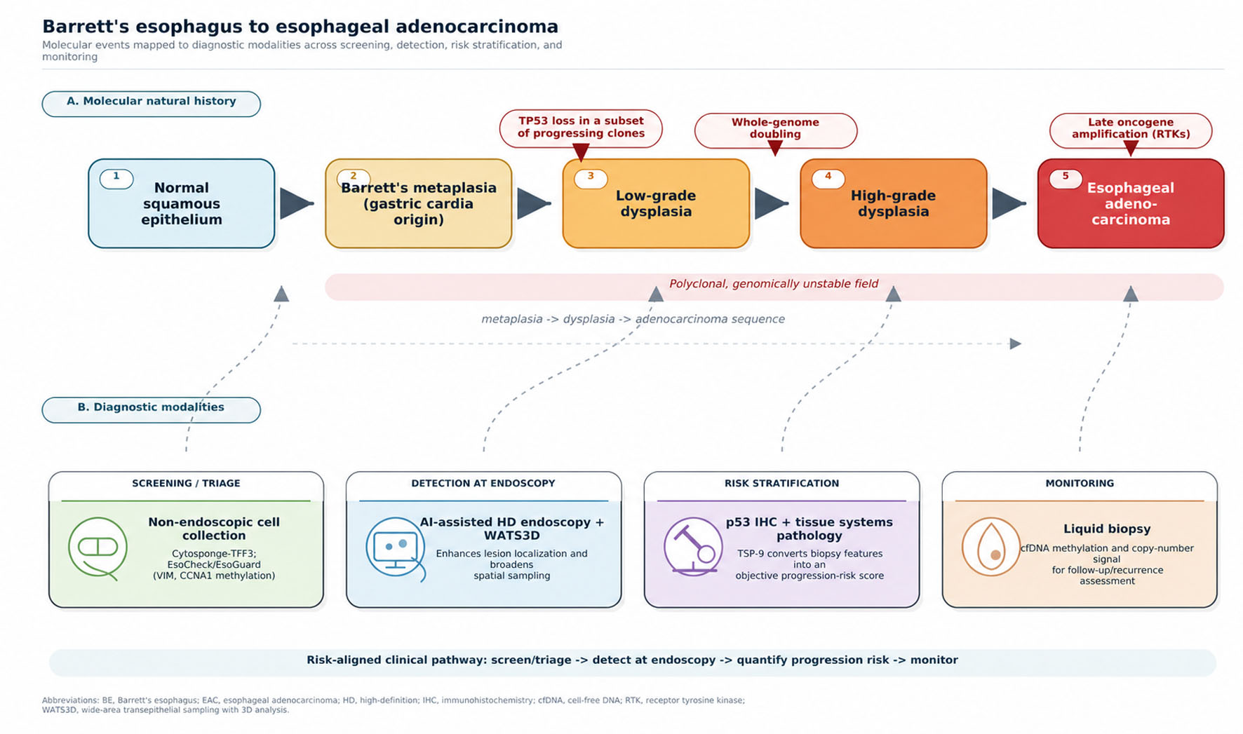
Esophageal adenocarcinoma (EAC) is among the most lethal gastrointestinal malignancies, and its incidence has risen markedly in Western populations over the last 50 years. Barrett’s esophagus (BE), the intestinal-type metaplasia that replaces the distal esophageal squamous mucosa, is the only known precursor of EAC and underpins current early-detection practice. Current surveillance—periodic endoscopy with random four-quadrant (Seattle protocol) biopsies graded for dysplasia—has two well-characterized limitations: forceps biopsies sample only a small portion of the metaplastic surface, and histopathological diagnosis of dysplasia, particularly low-grade dysplasia, has considerable interobserver variability. These limitations have prompted molecular approaches to risk stratification. This narrative review links the cellular and molecular pathobiology of the Barrett’s-to-EAC sequence with diagnostic approaches for risk stratification and early detection. We first summarize the genomic architecture of progression—TP53 loss and clonal expansion in a subset of progressing Barrett’s lesions, often preceding whole-genome doubling, together with epigenetic dysregulation and late oncogene amplification—along with evidence that BE is derived from gastric cardia progenitors at the single-cell level. We then review tissue-based adjuncts that provide objective information from the biopsy: p53 immunohistochemistry, multiplexed tissue systems pathology (the TSP-9/TissueCypher assay), and wide-area transepithelial sampling with computer-assisted three-dimensional analysis (WATS3D). We also appraise non-endoscopic molecular cell-collection devices used to triage patients for endoscopy: the Cytosponge coupled with trefoil factor 3, and the encapsulated balloon (EsoCheck) coupled with a methylated DNA panel (EsoGuard; methylated vimentin and cyclin A1). Finally, we consider liquid biopsy based on circulating cell-free DNA methylation, with artificial intelligence as a computational adjunct. Together, these modalities may convert the qualitative, observer-dependent question “Is there dysplasia?” into a quantitative assessment of an individual’s risk of progression. We examine where the evidence is strongest, where it remains preliminary, and how these tools could be combined into a risk-aligned surveillance model that detects EAC at a curable stage while reducing unnecessary procedures for patients at low risk.
Published
Issue
Section
License
Copyright (c) 2026 The authors

This work is licensed under a Creative Commons Attribution 4.0 International License.